What is Hip Dysplasia?
The hip joint is a ball (top of hip bone) and socket (in the pelvis) joint. Hip dysplasia is an abnormal developmental of the hip joint. This problem affects young growing animals and always results in the development of hip osteoarthritis.
What causes Hip Dysplasia?
This is a genetic (inherited) disorder that results from a complex combination of genes passed on from the patient’s parents. This combination of genes instructs the abnormal development of the joint, initially resulting in laxity (looseness) of the ball and socket joint. In other words, the ball moves in and out of the socket more than it should do, which results in abnormal loading of the soft bone and cartilage surfaces of both the ball and the socket. Both the looseness of the hip joint and / or the progressive development of osteoarthritis can result in pain and poor mobility.
What are the signs?
Hip dysplasia is a common condition, especially in large breed dogs. Not every patient who has hip dysplasia and develops hip osteoarthritis will become clinically affected; patients’ who are affected can have differences in severity of their problem and their age at which it becomes apparent. Key signs are lameness affecting the back legs and stiffness, with the latter most noticeable after rest following exercise. Difficulty rising and reluctance to jump or exercise are also reported features. Clinical signs tend to develop when the dog is immature and growing (five to ten months of age) where hip looseness and possibly osteoarthritis, are the source or hip pain. In adult dogs the hip osteoarthritis is the source pain.
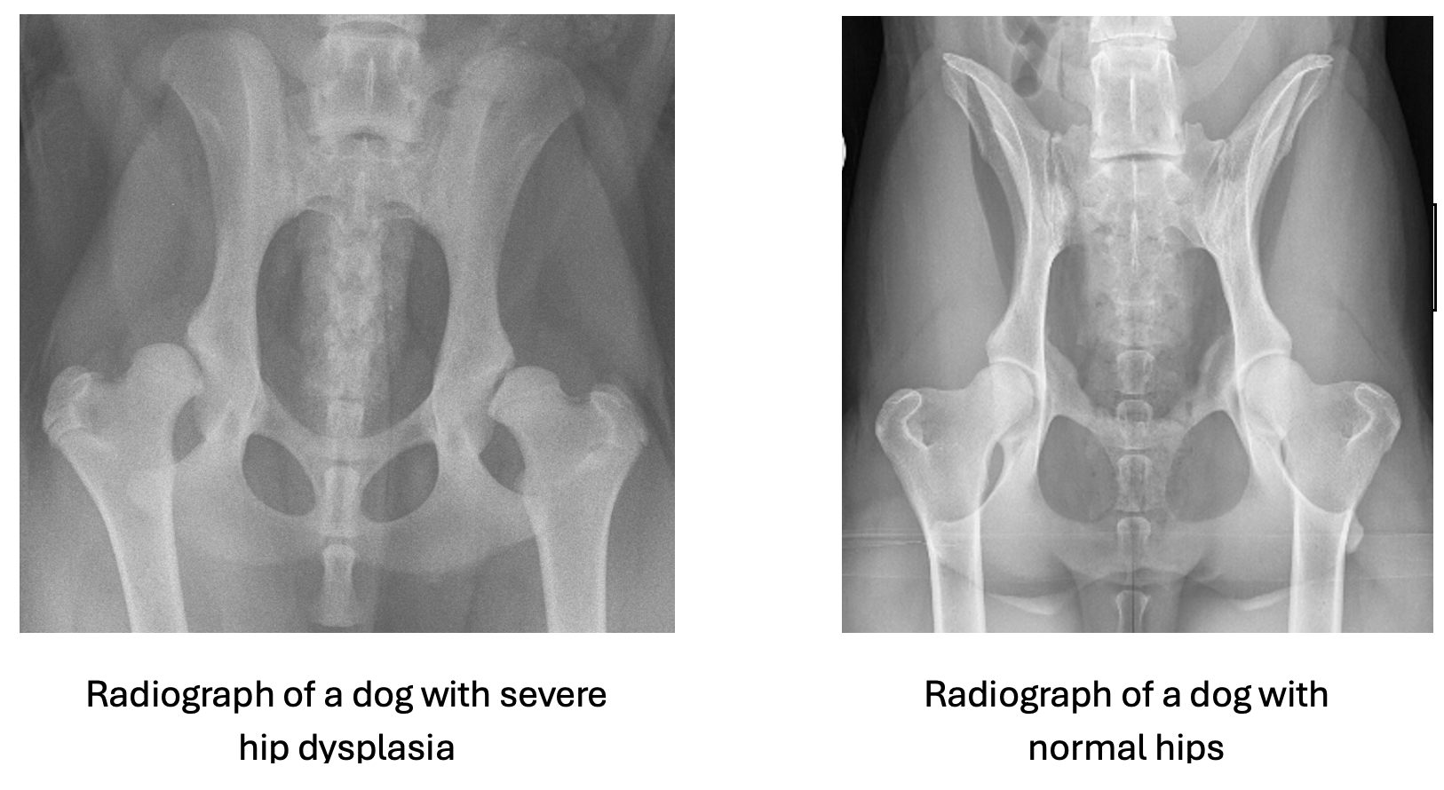
How is Hip Dysplasia diagnosed?
Examination of your pet and x-rays are required to confirm the diagnosis of Hip Dysplasia. X-rays then help confirm the diagnosis and severity of the problem including the severity of any associated osteoarthritis.
How is Hip Dysplasia treated?
Thankfully some patients can be managed satisfactorily without surgery. Non-surgical management involves a multi-faceted approach including bodyweight control, activity moderation and pain relief as the main stay. Some patients may also benefit from hydrotherapy and physiotherapy.
Surgical management is indicated in patients where non-surgical management is or becomes ineffective over the patient’s lifetime. Where hip dysplasia is severe, or osteoarthritis is present salvage surgery will be required.
Surgical management aimed at resolving hip pain and hip looseness can be considered in skeletally immature dogs, who meet specific criteria. This surgery requires the pelvis to be reshaped; it can be considered as corrective surgery. Unfortunately, not many dogs meet the criteria to be considered for this surgery.
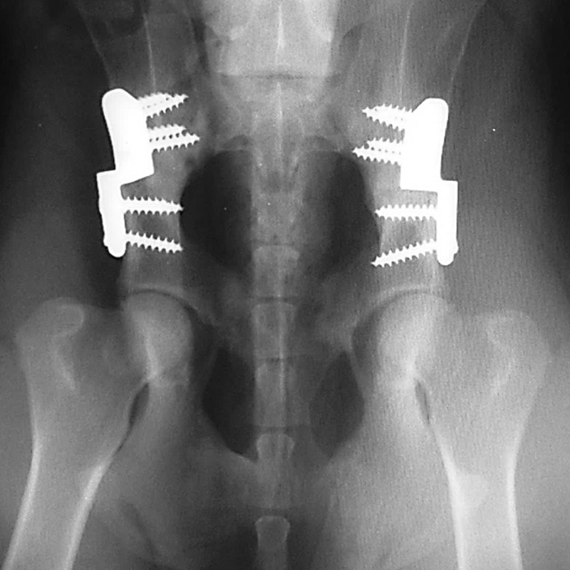
Corrective surgery: Pelvic osteotomy surgery requires the pelvis to be cut in either two or three places to allow the segment containing the socket to be rotated into a new position to capture the ball and improve the stability and fit of the hip joint. A metal bone plate and screws are then placed to hold the bones in this new position until the pelvis heals. This surgery preserves the dog’s own joint tissues and hopefully reduces the development / progression of osteoarthritis.

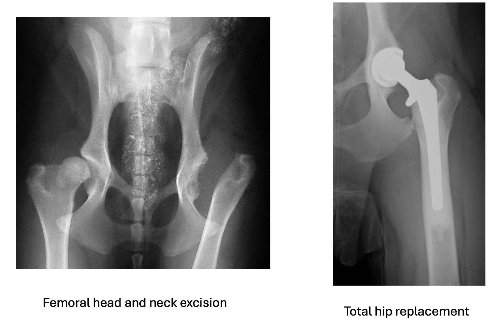
Salvage surgery – This will either be a total hip replacement or femoral head and neck excision.
Total hip replacement involves removing the painful hip joint and replacing it with an artificial (prosthetic) hip, consisting of a plastic socket and a metal head. This is a major surgical procedure but provides a rapid reduction in hip pain and resolution of lameness in most dogs. The recovery after total hip replacement is rapid and predictable and the functional outcome better in comparison to femoral head and neck excision. Whilst total hip replacement will most commonly be performed in medium to large breed dogs, the range of artificial hips available allows for it to be performed in small breed dogs and cats. Despite the excellent prognosis in most dogs’ complications can occur, most of which will require additional surgery.
Femoral head and neck excision involves removal of the femoral head and neck (ball); no artificial hip is placed. Over time a false joint will form; the limb is supported by the false joint scar tissue and the surrounding muscles. Although pain will be reduced, most patients will retain residual lameness, in part due to the reduction in hip joint motion and slight shortening of the limb. Recovery can be slow, and the outcome can be unpredictable. Small breed dogs and cats cope better than larger dogs after this surgery. Postoperative physiotherapy and hydrotherapy can play an important role in optimising recovery.