
Patellar luxation
What is patellar luxation?
The bony part at the front of your knee that you can feel is the patella, commonly known as the kneecap; it sits within the bottom part of the quadriceps muscle and a ligament attaches it to the top of the shin bone (tibia). The muscle, patella and the ligament make up the quadriceps mechanism. Patellar luxation is when the kneecap slips out of its cartilage lined groove (trochlea) at the bottom of the thigh bone (femur). The patella can luxate to the inner (medial) or the outer side (lateral) of its groove; occasionally it can luxate in both directions. Stability of the patella as the knee flexes and extends is important to allow the knee and leg to function properly. Patella luxation is more common in dogs than cats.
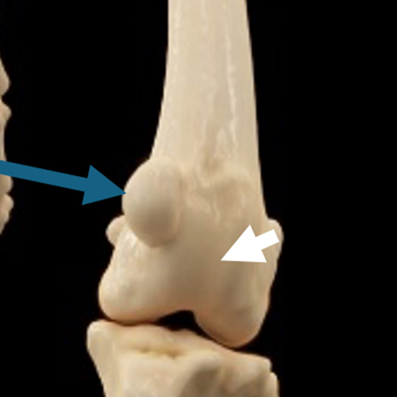
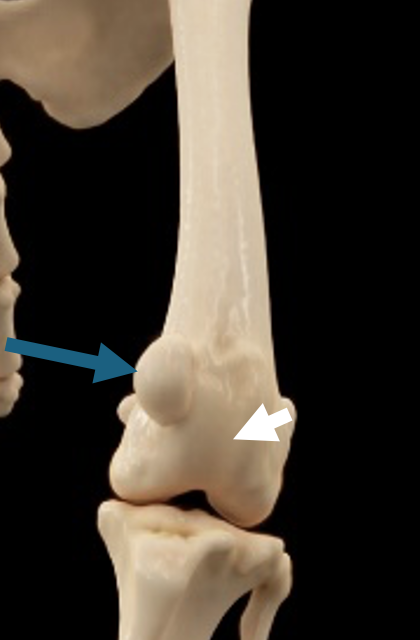
CT scan of a dog’s hind limb. The blue arrow indicates the luxated patella and white arrow the groove
What causes patellar luxation?
For most patients, patella luxation is a developmental problem, with genetic factors likely to be involved, although for some patient’s trauma may play a role. In most patients an underlying malalignment of the quadriceps mechanism and trochlear groove is present. Most commonly malalignment is due to deformity of the thigh or shin bone. Patella luxation frequently affects both hindlimbs, although this can be to differing degrees of severity.
What are the signs?
Dogs will often have an intermittent hop/skip on the affected limb when walking, which occurs as the patella luxates, and resolves once the patella relocates back into its groove. Alternatively, the affected limb may appear to collapse when the patella luxates. In some patients where the patella is permanently luxated a crouched back leg posture may be present. In more chronic cases or in more severely affected animals a hop/ skip may not be present, but a more persistent limp is. This is usually the result of chronic mal-tracking of the patella and with time, wear and tear (osteoarthritis) within the knee joint will develop. Patellar luxation can be present but may not be a cause of lameness in some patients.
How is patellar luxation diagnosed?
A diagnosis of both patellar luxation and its severity are made during your pet’s orthopaedic examination. As the severity increases any underlying bone deformity generally becomes more apparent. Although a diagnosis is achieved on examination, investigation involving imaging of the affected leg(s) is required. Based on the severity of the problem in the individual patient, your surgeon will advise as to whether x-rays or a CT scan will be required. Interpretation of this information, in combination with the examination findings, facilitates decision making as to which surgical procedures will be required to stabilise the patella in an individual patient.
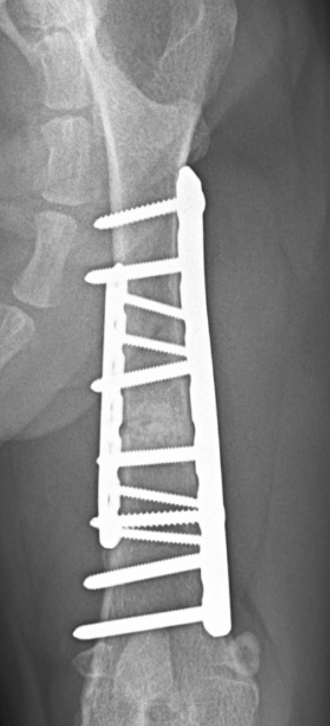
Corrective osteotomy of the femur bone
How is patellar luxation treated?
When surgery is required, the aim is to realign the quadriceps mechanism. For most patient’s a combination of procedures can be required. Most commonly surgery will involve:
- Patellar groove deepening – A block or wedge of bone is removed from the groove, retaining its cartilage covering. The bed from where this bone is removed is then deepened by removing more bone from within. The cartilage covered fragment of bone is then replaced into its original position but now sinks deeper into place. This provides a deeper home for the patella to sit in and restores the ‘walls’ of the groove which help to restrain it on either side.
- Tibial tuberosity transposition – This component of surgery shifts the attachment of the patellar ligament at the top of the shin bone to realign it with the patellar groove. The bone attachment of the ligament is cut, moved to its new position and stabilised using multiple pins and wire.
- Corrective osteotomy – This may be required in patients who have significant thigh and / or shin bone deformity. Corrective osteotomy involves breaking and reshaping the bone(s) in order to stabilise the patella. The broken and reshaped bone is stabilised using a bone plate and screws allowing it to heal. Additional procedures such as groove deepening or tibial tuberosity transposition may still be required.
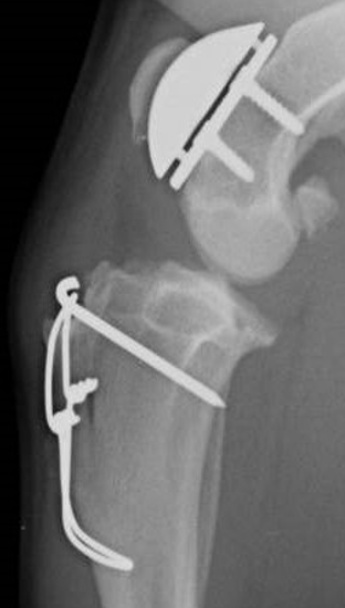
For patients where significant wear and tear of the surfaces of the patella and groove is present, pain and lameness can persist despite realignment and stabilisation of the patella. For these patients cutting out the damaged groove and replacing it with an artificial groove – patellar groove replacement – is the better surgical option. This may need to be performed with corrective osteotomy or tibial tuberosity transposition.
Patella groove replacement and tibial tuberosity transposition
How long is the recovery?
Most dogs will start walking on the operated leg within a few days of surgery with continued improvement over the following weeks. Your pet will receive painkillers for a few weeks after surgery. The recovery period following surgery is usually around 6-12 weeks to the point where the patient can be returned to normal activity levels; time scales will vary depending on the exact surgery which has been performed. Strict activity restrictions, which may include a period of pen / crate confinement, are imposed for the first 4-6 weeks. Full postoperative management and activity instructions will be provided and explained by the team when you collect your pet after their surgery. Initial checks on the wound and removal of any skin stitches will be undertaken by your own vet, however a follow up appointment will be arranged with your surgeon towards the end of the recovery period to ensure that your pet has made expected progress. At this follow up x-rays will be obtained, most commonly under sedation rather than anaesthesia to check the implants and to assess healing of the bone. If all is progressing as expected further rehabilitation guidelines will be given, and any further assessments scheduled. Unless indicated implants remain in place for life.
What is the outlook following surgery?
The outcome for most patients is good. All patients will develop osteoarthritis of the knee to some degree, but for most this is not a cause of pain or lameness. A small number of pets will retain some residual lameness and may require ongoing analgesic medication.
What are the potential complications following surgery?
Our surgeons will have a thorough discussion with you ahead of surgery to make sure you are fully informed regarding possible complications. If a complication develops your pet is likely to require assessment by your surgeon. Investigations including x-rays and knee joint fluid collection and analysis may be required. Complications are infrequent but can include such things as:
Infection – will require antibiotic medication ideally based on the results of sample analysis. For some patient’s infection may necessitate further surgery for it to be resolved.
Implant problems such as loosening or breakage – in some patients this may necessitate an additional procedure.
Recurrence of patella luxation – in some patients this may necessitate further surgery.


