Canine Cruciate Ligament Rupture
What is the cranial cruciate ligament?
The cranial cruciate ligament is one of the ligaments which provides stability to the dogs knee (stifle); the joint between the thigh bone (femur) and shin bone (tibia). The main job of the cranial cruciate ligament is to prevent forward movement of the shin bone relative to the thigh bone.
Why does the cranial cruciate ligament rupture?
In most dogs the cranial cruciate ligament becomes diseased, which results in its progressive degeneration and weakening over time, ultimately leading to its rupture, most often during a period of normal activity. The exact cause of this disease remains unclear, but genetics likely play a key role. Once the cranial cruciate ligament becomes diseased, osteoarthritis will develop in the affected knee. Traumatic rupture of a healthy cranial cruciate ligament occurs in a small number of dogs. These patients may present additional challenges because quite commonly these patients will have injuries to other knee ligaments.
What are the signs?
In the early stages of cranial cruciate ligament disease, generally before knee instability becomes apparent, signs can be minimal e.g. intermittent lameness and stiffness after rest which resolve with activity. Over time, as the cranial cruciate ligament continues to degenerate, abnormal movement (instability) of the knee develops as the ligament begins to fray and ultimately ruptures. Lameness and dysfunction of the affected leg become more obvious, with some dogs refusing to bear weight on the leg. The knee will become thickened, and muscle will be lost on the affected leg. Some dogs will experience cranial cruciate ligament disease / rupture in both back legs in quick succession. These dogs may still be able to walk albeit with very poor back leg use, however some may be unable to stand on their back legs.
How is cranial cruciate ligament rupture diagnosed?
Diagnosing cranial cruciate ligament rupture relies on thorough examination of the knee and x-rays. Sedation or general anaesthesia is required for these procedures. Diagnosis of cranial cruciate ligament rupture is made by identifying instability of the knee joint. X-rays allow the severity of arthritis to be assessed, to exclude other possible knee conditions and to enable surgical planning, to ensure that the best management option is selected for each individual patient. Collection of knee joint fluid may also be required to exclude other possible knee problems. In the small number of dogs where there is no appreciable knee instability apparent on examination, visual inspection of the ligament may be required to confirm the diagnosis. This can be achieved either by making a small surgical incision (arthrotomy) into the knee or by using ‘keyhole surgery’ with a camera (arthroscopy).

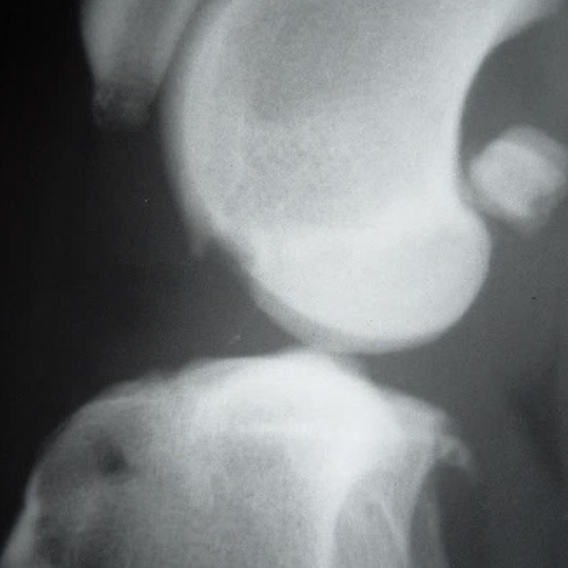
An x-ray of a dogs knee. The arrows indicate the osteoarthritic bone which has developed as a consequence of cranial cruciate ligament rupture
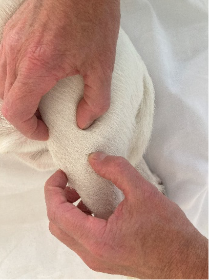
The knee is examined for instability using the cranial draw test
So, my dog’s ruptured its cranial cruciate ligament, what now?
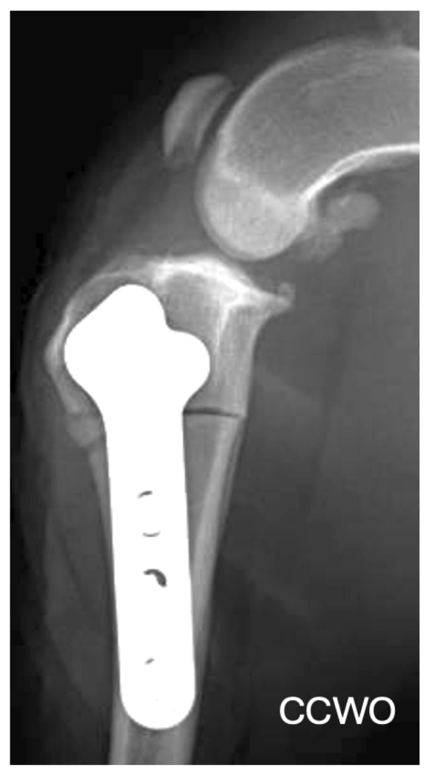
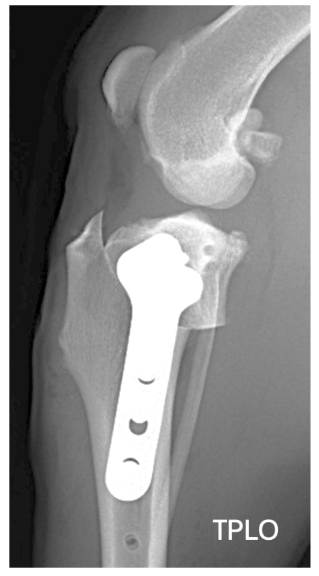
Non-surgical management is extremely unlikely to significantly improve lameness and return dogs to their previous level of activity and comfort. Very small dogs may stand a better chance of improving without surgery, but even so, this can take a long time and rarely will they make a full recovery. Surgery is the management of choice for cranial cruciate ligament rupture. Surgical options include either ligament replacement technique or a bone cutting (osteotomy) technique. Both techniques address the knee instability albeit by different means. Osteotomy surgeries include tibial plateau levelling osteotomy (TPLO) and cranial closing wedge osteotomy (CCWO) and are our surgical techniques of choice given their good prognosis for improving limb function and resorting active pain free mobility. In most dogs the surgeon will also look inside the knee, in particular to check the knee shock absorbers (meniscus), which can be damaged because of knee instability. If present the damaged portion of the meniscus will be removed.