
Humeral Intracondylar Fissure
Where is the humerus bone and what is a humeral intracondylar fissure?
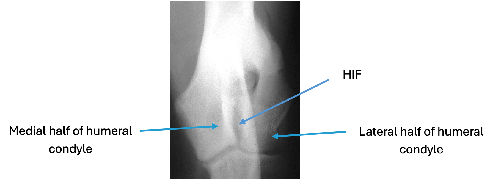
The humerus is a large bone in the front leg. Along with the scapula (shoulder blade) it forms the shoulder joint at the top and along with the radius and ulna (forearm) bones it makes up the elbow joint at the bottom. The bottom part of the humerus which fits together with the radius and ulna bones is called the humeral condyle. A humeral intracondylar fissure (HIF) is a gap or crack which is present in the middle of the humeral condyle. This gap or crack may extend part or all the way through the humeral condyle, potentially separating the inside (medial) and outside (lateral) halves.
What causes a HIF to develop?
The exact reason for HIF remains uncertain. The condition is most commonly seen in Spaniels, particularly English Springer Spaniels, and as such genetics are likely to play a role, as may front leg conformation and / or possibly poor fitting of the elbow joint. There are distinct groups of dogs affected by HIF. Firstly, in skeletally immature patients an incomplete ossification of the humeral condyle (IOHC) may be present. For many years it was assumed that IOHC was the cause even when HIF was identified in adult dogs, however, a HIF can develop in a humeral condyle previously identified to have been normal. In this group of dogs HIF is now considered a stress fracture.
What are the signs?
When HIF is present lameness can be variable, being mild and intermittent for some and marked and persistent for others. Once lameness develops it tends to be progressive in nature. The weakness that HIF causes in the humeral condyle can result in some dogs fracturing their humeral condyle; this can happen during periods of normal activity. Fractures are more likely to involve one portion of the humeral condyle. These uni-condylar fractures most frequently affect the lateral (outer) half rather than the medial (inner) half. In some dogs both halves of the humeral condyle fracture. These di-condylar fractures tend to have a ‘Y’ or ‘T’ shaped pattern. Y and T fractures are some of the most difficult fractures we repair. HIF commonly affects both elbows; not necessarily at the same time or to the same extent.
How is a HIF diagnosed?
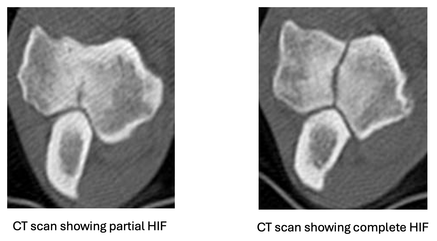
Examination of the patient will usually identify pain on manipulation of the elbow and often reduced muscling of the affected limb. Larger fissures might be identifiable with x-rays, however for many dogs x-rays will fail to identify HIF, even when it is present. Computed tomography (CT) is performed to allow for a more accurate assessment of the humeral condyle.
So, my pet has a HIF, what now?
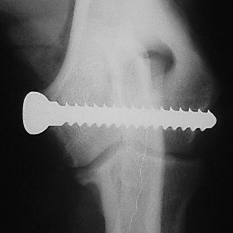
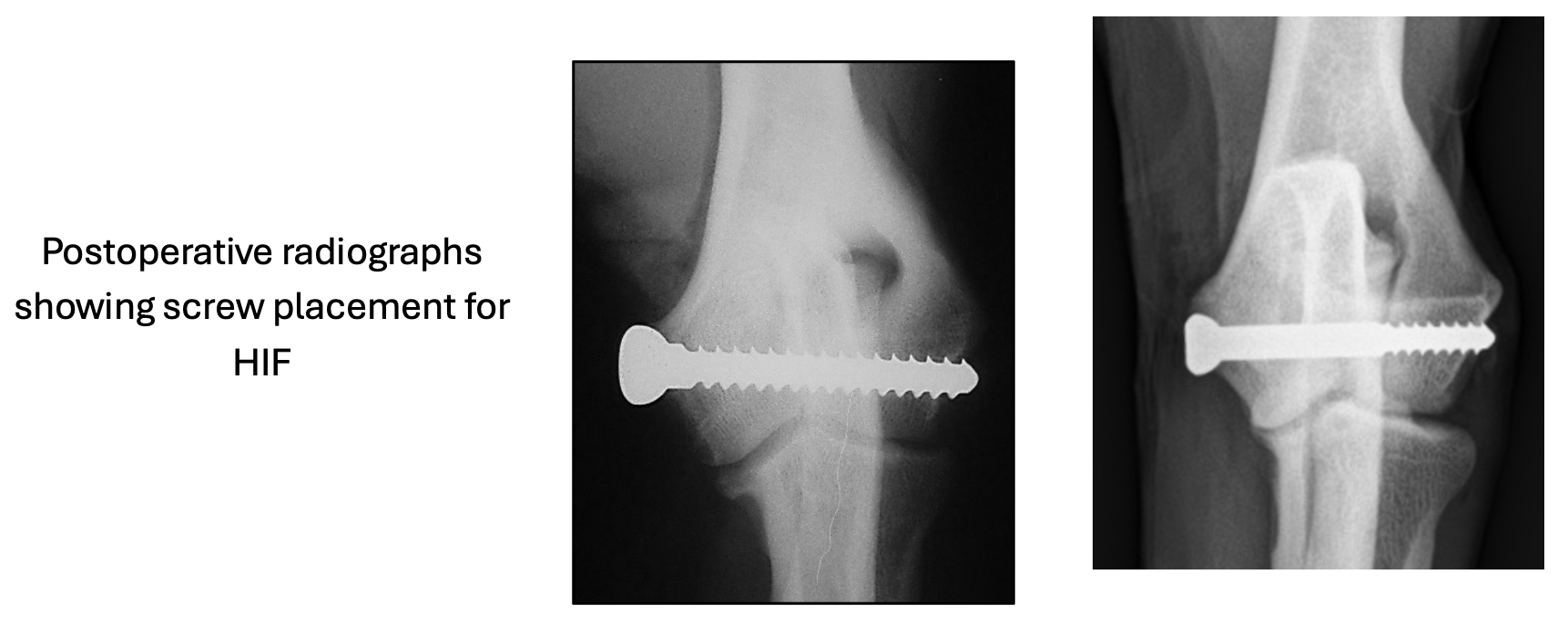
In some patients HIF (either partial or complete) may be identified on either x-ray or CT, but before it has started to cause elbow discomfort and lameness. If this is the case, your surgeon will discuss options for management moving forward. When a HIF is the cause of lameness surgery is recommended. The rationale for surgery is twofold; firstly to resolve / reduce elbow pain and lameness and secondly to reduce the possibility of a humeral condylar fracture developing. For most patients’ surgery involves placing a large bone screw across the humeral condyle to restore its stability and strength. Accurate placement of a large screw within the humeral condyle is imperative; as such accurate screw placement can be facilitated using a guide or intra-operative x-rays (fluoroscopy).
How long is the recovery?
Most dogs will use the operated leg within a few hours of surgery with continued progress over the coming weeks. Your pet will receive painkillers for a few days after surgery. The recovery period following screw placement for HIF is usually around 3 – 4 weeks to the point where the patient can be returned to normal activity levels. Activity restrictions are imposed for the first 2-3 weeks. Full postoperative management and activity instructions will be provided and explained by the team when you collect your pet after their surgery. Initial checks on the wound and removal of any skin stitches will be undertaken by your own vet, however a follow up appointment will be arranged with your surgeon towards the end of the recovery period to ensure that your pet has made expected progress. At this follow up appointment x-rays will generally only be obtained if indicated. If all is progressing as expected further rehabilitation guidelines will be given, and any further assessments scheduled. Unless indicated implants remain in place for life.
What is the outlook following surgery?
Most dogs will have a good long-term functional outcome; some will retain residual lameness and may require analgesic medication.
What are the potential complications following surgery?
Our surgeons will have a thorough discussion with you ahead of surgery to make sure you are fully informed regarding possible complications. If a complication develops your pet is likely to require assessment by your surgeon. Investigations including x-rays and elbow joint fluid collection and analysis may be required. Complications include such things as:
Infection – will require antibiotic medication ideally based on the results of sample analysis. For some patients’ infection may necessitate further surgery as part of their management.
Screw problems such as loosening or breakage – These are uncommon complications. There can be several different reasons for implant problems developing. Most implant problems will necessitate further surgery. We do not expect every HIF to heal. As such there is a small risk of future screw failure and humeral condylar fracture.
Wound problems – Whilst these can be seen following any surgery, the risk is small. A very small number of dogs may develop a fluid swelling (seroma) at the wound site; this will tend to resolve over time without intervention.


