
Elbow dysplasia
What is elbow dysplasia?
Elbow dysplasia is an abnormal development of the elbow; the joint made up of the bottom of the funny bone (humerus) and the bones of the forearm (radius & ulna). Elbow dysplasia is a complex disease, it encompasses different conditions which although often considered as separate problems, share common consequences of cartilage damage, osteoarthritis development, elbow pain and lameness. Once osteoarthritis develops it will continue to progress in severity over the dog’s lifetime. Elbow osteoarthritis is one of the most challenging conditions to manage effectively.
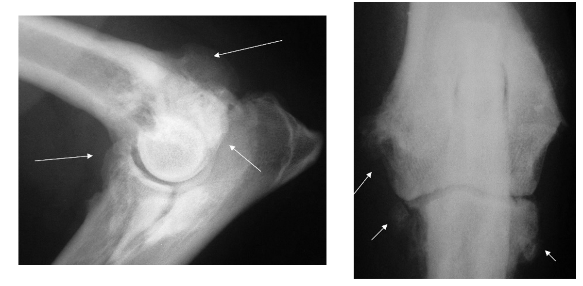
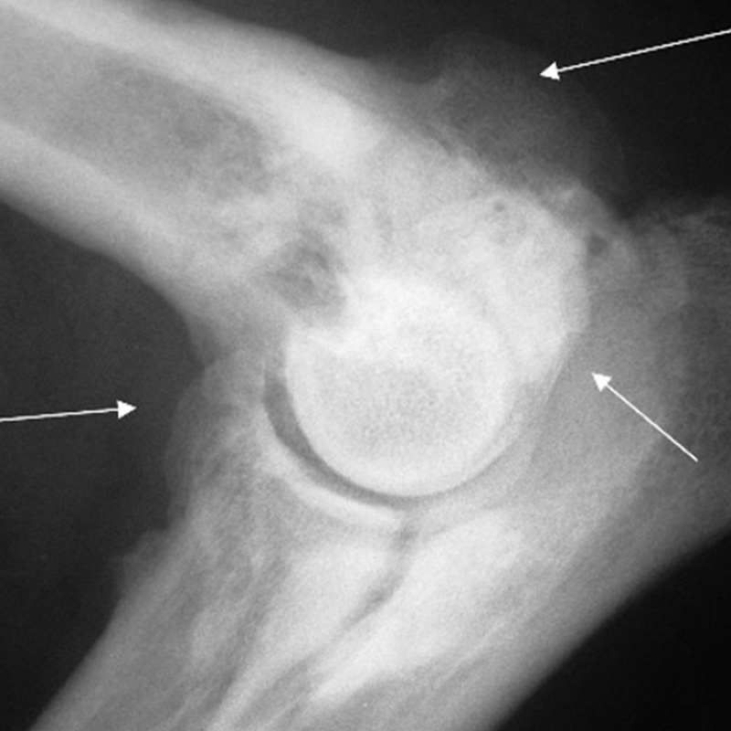
Elbow x-rays from a 10-year-old labrador who has severe elbow osteoarthritis. The white arrows indicate the osteoarthritic bone that has developed. This dog had medial coronoid process disease diagnosed at 8 months of age.
The most common condition within the elbow dysplasia complex is medial coronoid process disease (MCPD); osteochondritis dissecans (OCD) and ununited anconeal process are less common, whilst poor fitting (incongruity) of the elbow joint can also be present. These conditions may be present concomitantly, adding to the complexity of the problem.
What causes elbow dysplasia?
Elbow dysplasia is genetic condition. Genes inherited from the dog’s parents, most likely influenced by environmental factors contribute to the complexity of the disease making accurate identification of the cause of the disease extremely difficult. Health schemes are available e.g. BVA Elbow Dysplasia Scheme for Dogs, which breeders are encouraged to utilise. The scheme aims to remove the highest risk individuals from the breeding population to hopefully reduce the prevalence of this debilitating disease over time.
What are the signs? Forelimb lameness is the most common sign of elbow dysplasia. The severity of lameness can vary considerably between patients. It is not uncommon for both elbows to be affected and many dogs will be lame in both forelimbs. The age at which dogs develop lameness can vary depending on their elbow condition, however the majority will show signs of lameness between 6-18 months of age. Some dogs may not show any clinical signs until they reach adulthood.
How is elbow dysplasia diagnosed?
Thorough orthopaedic examination by one of our experienced orthopaedic surgeons will localise pain to the elbow joint(s); alteration in elbow joint range of motion, fluid swelling (joint effusion) and thickening of the joint(s) may also be identifiable.
Your local vet may have already acquired elbow x-rays, and these may show changes such as osteoarthritis. Whilst useful, x-rays are limited in the information they can provide, and computed tomography (CT) is therefore recommended to more thoroughly assess the elbow joint. Arthroscopy (keyhole surgery) also allows for thorough assessment of the joint, in particular the health of the cartilage surfaces, and also offers and opportunity for arthroscopic surgery to be performed if indicated.
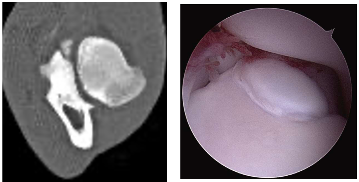
CT and arthroscopic images from a dog with medial coronoid process disease. In this dog the medial coronoid process was fragmented. The fragment was removed arthroscopically
How is elbow dysplasia treated?
It is important for you to know that elbow dysplasia is an incurable disease, however there are many management strategies which can be utilised in an attempt to resolve or reduce lameness and improve the mobility of your dog. Your surgeon will consider all the information obtained from your dog’s history, orthopaedic examination and imaging findings, when deciding on the most appropriate management strategy for your dog.
Management options can include such things as:
- Non-surgical (medical) management – involves a multi-faceted approach including bodyweight control, activity moderation and pain relief as the main stay. Some patients may also benefit from hydrotherapy and physiotherapy.
- Elbow arthroscopy – allows joint inspection and removal of loose bone / cartilage fragments
- Ulna osteotomy – cutting the ulna bone to manage poor fitting (incongruity) of the elbow joint.
- Screw placement and ulna osteotomy for ununited anconeal process
- Salvage surgery – elbow arthrodesis or elbow replacement for the end stage osteoarthritic joint. These procedures are rarely performed.
The prognosis associated with management of elbow dysplasia is variable between patients. Irrespective of what management is undertaken we expect osteoarthritis to develop and progress over the patient’s lifetime. What cannot be predicted is how much osteoarthritis will develop and how it will impact it on your dog’s mobility.


